
A virtual reconstruction of the deformed DFN3-150 Early Pleistocene Paradolichopithecus cranium from Dafnero-3, Greece = Ψηφιακή ανακατασκευή του παραμορφωμένου κρανίου DFN3-150 του κάτω-πλειστοκαινικού Paradolichopithecus από τη θέση Δαφνερό-3, Ελλάδα.
Περίληψη
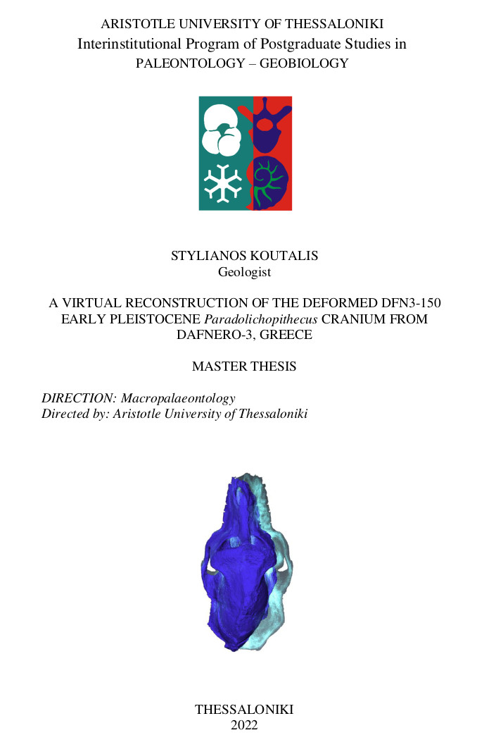
Η παρούσα εργασία ασχολείται με την ψηφιακή ανακατασκευή του παραμορφωμένου κρανίου LGPUT DFN3-150 του Κάτω-Πλειστοκαινικού Paradolichopithecus aff. αrvernensis της θέσης Δαφνερό-3, Ελλάδα. Η αποσπασματική παρουσία του Paradolichopithecus στην Ευρασία στο Κάτω Πλειστόκαινο και η απότομη εξαφάνιση του από το αρχείο απολιθωμάτων στα τέλη του Κάτω Πλειστοκαίνου, τοποθετεί το παρόν κρανίο στο κέντρο του παλαιοντολογικού ενδιαφέροντος, τόσο σε ταξινομικό όσο και εξελικτικό επίπεδο. Έχουν περάσει περισσότερα από εκατό χρόνια από όταν ο Wilhelm C. R?ntgen εισήγαγε τις ακτίνες Χ στο κοινό, και λιγότερο από μια γενιά που η αξονική τομογραφία αξιοποιήθηκε στην ανακατασκευή απολιθωμένων κρανίων. Παρέχεται μια σύντομη περιγραφή της ιστορίας, των αρχών και των μηχανισμών που επιτρέπουν στο εργασιακό οργανόγραμμα να αποτελεί ένα καλώς διαμορφωμένο πλαίσιο έρευνας. Αξιοποιώντας δύο βασικές αρχές της βιολογίας σκελετικών δειγμάτων στην παρούσα εργασία, την πλευρική αμφισυμμετρία και την αρχικά ομαλή ακολουθία καμπύλωσης των εξωτερικών επιφανειών των κρανίων, το LGPUT DFN3-150 υποβλήθηκε αρχικά στην διαδικασία της «κατάτμησης», στο υπολογιστικό περιβάλλον των εφαρμογών Aviso® 9.2.0 Lite και Aviso® 7.0.0 (Visualization Sciences Group). Η διαδικασία αυτή εφαρμόζεται απευθείας στα δεδομένα της αξονικής τομογραφίας, επιτρέποντας τον διαχωρισμό του ιζήματος από τα οστά, όπως επίσης την διαίρεση του οστού σε διαφορετικές ομάδες. Οι ομάδες θα μπορέσουν στη συνέχεια να διαχειριστούν αυτόνομα, ώστε οποιαδήποτε δομή να μπορεί να ανακατασκευαστεί επανατοποθετώντας τα επιμέρους τμήματά της. Για το LGPUT DFN3-150 διαχωρίστηκαν είκοσι πέντε τμήματα από το αρχικό απολίθωμα, αλλά δεν αποτελούν όλα προϊόντα πλήρους διάρρηξης. Τα περισσότερα από αυτά σχεδιάστηκαν και «αποσπάσθηκαν» ψηφιακά μεταξύ διατηρημένων ραφών, ή ακολουθώντας κάποιες ζώνες ραφών. Για να μειωθεί η διαγενετική συν- και μετά-ταφική παραμόρφωση, ενώ παράλληλα να μελετηθούν τα αποτελέσματα εφαρμογής διαφορετικών τακτικών ανακατασκευής, δημιουργήθηκαν τρία μοντέλα. Τα πρώτα δύο αναφέρονται στην προσπάθεια ελαχιστοποίησης της παραμόρφωσης στη δεξιά πλευρά του κρανίου, διορθώνοντας την μετατόπιση που υπέστη η δεξιά άνω γνάθος. Το τρίτο μοντέλο αποτελεί ένα μοντέλο «αντικατοπτρισμού» όπου όλα τα θραύσματα της αριστερής πλευράς αντιγράφηκαν και αναστράφηκαν. Έπειτα, ακολουθώντας την επανατοποθέτηση του αριστερό μετωπικού θραύσματος με το αντίγραφό του, σε ένα οβελιαίο επίπεδο συμμετρίας, τα υπόλοιπα θραύσματα επανατοποθετήθηκαν ακολουθώντας την θέση τους στο άλλα δύο μοντέλα. Όταν αυτά τα μοντέλα αντιπαραβάλλονται με το αρχικό απολίθωμα, η εντονότερη διαφορά έγκειται στο νέο διευρημένο πλάτος. Αυτό επηρεάζει το σχήμα του ινιακού τρήμματος, προσδίδοντάς του ένα πιο κυκλικό περίγραμμα, χαρακτήρας που παρατηρείται και στα τρία μοντέλα. Σχετικά με την παραμόρφωση μεταξύ του αρχικού απολιθώματος και του μοντέλου αντικατοπτρισμού, το τελευταίο διαφοροποιείται στο σχήμα της υπερόφριας ακρολοφίας, το καμπύλο περίγραμμα της οποίας συνιστά ένα θέμα προς συζήτηση, κατά πόσο δηλαδή αποτελεί αυθεντικό μορφολογικό χαρακτήρα ή είναι το προϊόν της πλαστικής παραμόρφωσης που δεν μπορούσε να εξαλειφθεί με τις παρούσες τεχνικές. Καταλήγοντας, μια ενδελεχής εφαρμογή μεθόδων «αντιστροφής» της παραμόρφωσης θα ήταν χρήσιμη για την εξαγωγή ενός πλήρως ανακατασκευασμένου μοντέλο του LGPUT DFN3-150 καθώς η πλαστική παραμόρφωση ευθύνεται για την πλειονότητα της συν- και μετά-ταφικής επίδρασης στο απολίθωμα.
Πλήρες Κείμενο:
PDFΑναφορές
Alba, D.M., Delson, E., Carnevale, G., Colombero, S., Delfino, M., Giuntelli, P., Pavia, M., Pavia, G., 2014. First joint record of Mesopithecus and cf. Macaca in the Miocene of Europe. J. Hum. Evol. 67, 1–18. https://doi.org/10.1016/j.jhevol.2013.11.001
Alba, D.M., Delson, E., Morales, J., Montoya, P., Romero, G., 2018. Macaque remains from the early Pliocene of the Iberian Peninsula. J. Hum. Evol. 123, 141–147. https://doi.org/10.1016/j.jhevol.2018.07.005
Antoniades, V., Penolidis, T., Dardiotis, G., 2009. History of ancient philosophy (1st ed.) Athens: Crateros. ISBN 978-960-89886-1-3 (in greek).
Amano, H., Kikuchi, T., Morita, Y., Kondo, O., Suzuki, H., Ponce de León, M.S., Zollikofer, C.P.E., Bastir, M., Stringer, C., Ogihara, N., 2015. Virtual reconstruction of the Neanderthal Amud 1 cranium: VIRTUAL RECONSTRUCTION OF AMUD 1 CRANIUM. Am. J. Phys. Anthropol. 158, 185–197. https://doi.org/10.1002/ajpa.22777
Amiez, C., Sallet, J., Hopkins, W.D., Meguerditchian, A., Hadj-Bouziane, F., Ben Hamed, S., Wilson, C.R.E., Procyk, E., Petrides, M., 2019. Sulcal organization in the medial frontal cortex provides insights into primate brain evolution. Nat. Commun. 10, 3437. https://doi.org/10.1038/s41467-019-11347-x
Atkinson, E.G., Rogers, J., Mahaney, M.C., Cox, L.A., Cheverud, J.M., 2015. Cortical Folding of the Primate Brain: An Interdisciplinary Examination of the Genetic Architecture, Modularity, and Evolvability of a Significant Neurological Trait in Pedigreed Baboons (Genus Papio ). Genetics 200, 651–665. https://doi.org/10.1534/genetics.114.173443
Babalola, A.M., 2019. Visualization of Voxel Volume Emission and Absorption of Light in Medical Biology. Curr. Trends Biostat. Biom. 1. https://doi.org/10.32474/CTBB.2019.01.000114
Bagley, B., Sastry, S.P., Whitaker, R.T., 2016. A Marching-tetrahedra Algorithm for Feature-preserving Meshing of Piecewise-smooth Implicit Surfaces. Procedia Eng. 163, 162–174. https://doi.org/10.1016/j.proeng.2016.11.042
Balzeau, A., Grimaud-Hervé, D., Détroit, F., Holloway, R.L., Combès, B., Prima, S., 2013. First description of the Cro-Magnon 1 endocast and study of brain variation and evolution in anatomically modern Homo sapiens. Bull. Mém. Société Anthropol. Paris 25, 1–18. https://doi.org/10.1007/s13219-012-0069-z
Barequet, G., Vaxman, A., 2008. Nonlinear Interpolation between Slices. Int. J. Shape Model. 14, 39–60.
Beaudet, A., Dumoncel, J., de Beer, F., Duployer, B., Durrleman, S., Gilissen, E., Hoffman, J., Tenailleau, C., Thackeray, J.F., Braga, J., 2016. Morphoarchitectural variation in South African fossil cercopithecoid endocasts. J. Hum. Evol. 101, 65–78. https://doi.org/10.1016/j.jhevol.2016.09.003
Beaudet, A., Gilissen, E., 2018. Fossil Primate Endocasts: Perspectives from Advanced Imaging Techniques, in: Bruner, E., Ogihara, N., Tanabe, H.C. (Eds.), Digital Endocasts. Springer Japan, Tokyo, pp. 47–58. https://doi.org/10.1007/978-4-431-56582-6_4
Belmaker, M., 2010. The presence of a large cercopithecine (cf. Theropithecus sp.) in the •Ubeidiya formation (Early Pleistocene, Israel). J. Hum. Evol. 11.
Benammi, M., Aidona, E., Merceron, G., Koufos, G.D., Kostopoulos, D.S., 2020. Magnetostratigraphy and Chronology of the Lower Pleistocene Primate Bearing Dafnero Fossil Site, N. Greece. Quaternary 3, 22. https://doi.org/10.3390/quat3030022
Benazzi, S., Bookstein, F.L., Strait, D.S., Weber, G.W., 2011. A new OH5 reconstruction with an assessment of its uncertainty. J. Hum. Evol. 61, 75–88. https://doi.org/10.1016/j.jhevol.2011.02.005
Berger, M., Yang, Q., Maier, A., 2018. X-ray Imaging, in: Maier, A., Steidl, S., Christlein, V., Hornegger, J. (Eds.), Medical Imaging Systems, Lecture Notes in Computer Science. Springer International Publishing, Cham, pp. 119–145. https://doi.org/10.1007/978-3-319-96520-8_7
Bookstein, F.L., 1991. Morphometric Tools for Landmark Data: Geometry and Biology. Cambridge University Press, Cambridge
Bosma, M., 2000. Iso-Surface Volume Rendering: speed and accuracy for medical applications. Ph.D. Thesis, University of Twente, 142pp.
Bosman, A.M., Buck, L.T., Centeno, H.R., Lahr, M.M., Stringer, C., Harvati, K., 2019. Modern human origins and dispersal, Words, bones, genes, tools. Kerns Verlag, Tübingen.
Bouchet, F., Ribéron, A., Heaton, J.L., 2019. The inner craniodental anatomy of the Papio specimen U.W. 88-886 from the Early Pleistocene site of Malapa, Gauteng, South Africa 15.
Chung, B.S., Jeon, C.-Y., Huh, J.-W., Jeong, K.-J., Har, D., Kwack, K.-S., Park, J.S., 2019. Rise of the Visible Monkey: Sectioned Images of Rhesus Monkey. J. Korean Med. Sci. 34, e66. https://doi.org/10.3346/jkms.2019.34.e66
Cignoni, P., Montani, C., Scopigno, R., Puppo, E., 2005. Optimal Isosurface Extraction, in: Visualization Handbook. Elsevier, pp. 69–82. https://doi.org/10.1016/B978-012387582-2/50006-X
Cirilli, O., Melchionna, M., Serio, C., Bernor, R.L., Bukhsianidze, M., Lordkipanidze, D., Rook, L., Profico, A., Raia, P., 2020. Target Deformation of the Equus stenonis Holotype Skull: A Virtual Reconstruction. Front. Earth Sci. 8, 247. https://doi.org/10.3389/feart.2020.00247
Clark, D.P., Badea, C.T., 2014. Micro-CT of rodents: State-of-the-art and future perspectives. Phys. Med. 30, 619–634. https://doi.org/10.1016/j.ejmp.2014.05.011
Connolly, C.J., 1950. The external morphology of the primate brain. CC Thomas, Springfield
Conroy, G.C., Vannier, M.W., 1984. Noninvasive Three-Dimensional Computer Imaging of Matrix-Filled Fossil Skulls by High-Resolution Computed Tomography. Science 226, 456–458. https://doi.org/10.1126/science.226.4673.456
Custodio, L., Pesco, S., Silva, C., 2019. An extended triangulation to the Marching Cubes 33 algorithm. J. Braz. Comput. Soc. 25, 6. https://doi.org/10.1186/s13173-019-0086-6
Delson, E., Nicolaescu-Plopsor, D., 1975. Paradolichopithecus, a large terrestrial monkey (Cercopithecidae, Primates) from the Plio-Pleistocene of southern Europe and its importance for mammalian biochronology. VIth Congress Regional Committee of Mediterranean Neogene Stratigraphy, Bratislava, pp. 91-96.
Depéret, C., 1929. Dolichopithecus arvernensis Depéret. Nouveau singe du Pliocène supérieur de Sénèze (Haute-Loire). Travaux et Documents des Laboratoires de Géologie de Lyon 12, 5-12
De Vos, J., Van der Made, J., Athanassiou, A., Lyras, G., Sondaar, P. Y., and Dermitzakis, M. D., (2002). Preliminary note on the late Pliocene fauna from Vatera (Lesvos, Greece). In Annales géologiques des Pays helléniques (Vol. 39, No. 3).
Di Vincenzo, F., Profico, A., Bernardini, F., Cerroni, V., Dreossi, D., Schlager, S., Zaio, P., Benazzi, S., Biddittu, I., Rubini, M., Tuniz, C., Manzi, G., 2017. Digital reconstruction of the Ceprano calvarium (Italy), and implications for its interpretation. Sci. Rep. 7, 13974. https://doi.org/10.1038/s41598-017-14437-2
Dumoncel, J., Subsol, G., Durrleman, S., Bertrand, A., Jager, E., Oettlé, A.C., Lockhat, Z., Suleman, F.E., Beaudet, A., 2021. Are endocasts reliable proxies for brains? A 3D quantitative comparison of the extant human brain and endocast. J. Anat. 238, 480–488. https://doi.org/10.1111/joa.13318
El Albani, A., Mangano, M.G., Buatois, L.A., Bengtson, S., Riboulleau, A., Bekker, A., Konhauser, K., Lyons, T., Rollion-Bard, C., Bankole, O., Lekele Baghekema, S.G., Meunier, A., Trentesaux, A., Mazurier, A., Aubineau, J., Laforest, C., Fontaine, C., Recourt, P., Chi Fru, E., Macchiarelli, R., Reynaud, J.Y., Gauthier-Lafaye, F.,
Canfield, D.E., 2019. Organism motility in an oxygenated shallow-marine environment 2.1 billion years ago. Proc. Natl. Acad. Sci. 116, 3431–3436. https://doi.org/10.1073/pnas.1815721116
Elliott, J.C., Dover, S.D., 1982. X-ray microtomography. J. Microsc. 126, 211–213. https://doi.org/10.1111/j.1365-2818.1982.tb00376.x
Elton, S., 2014. Macaques at the margins: the biogeography and extinction of Macaca sylvanus in Europe 14.
Erickson, G.M., 2005. Assessing dinosaur growth patterns: a microscopic revolution. Trends Ecol. Evol. 20, 677–684. https://doi.org/10.1016/j.tree.2005.08.012
Eronen, J.T., Rook, L., 2004. The Mio-Pliocene European primate fossil record: dynamics and habitat tracking. J. Hum. Evol. 47, 323–341. https://doi.org/10.1016/j.jhevol.2004.08.003
Falconer, I., 1997. J J Thomson and the discovery of the electron. IOP Publ. Ltd, Physics Education 32, 7. https://doi.org/10.1088/0031-9120/32/4/015
Falk, D., 2015. Evolution of the Primate Brain, in: Henke, W., Tattersall, I. (Eds.), Handbook of Paleoanthropology. Springer Berlin Heidelberg, Berlin, Heidelberg, pp. 1495–1525. https://doi.org/10.1007/978-3-642-39979-4_37
Falk, D., 2012. Chapter 12 - Hominin paleoneurology: Where are we now? 18.
Falk, D., 1981. Sulcal patterns of fossil Theropithecus baboons: Phylogenetic and functional implications. Int. J. Primatol. 2, 57–69. https://doi.org/10.1007/BF02692300
Falk, D., 1978. Brain evolution in old World monkeys. Am. J. Phys. Anthropol. 48, 315–319. https://doi.org/10.1002/ajpa.1330480307
Fleagle, J.G., 2013. Primate adaptation and evolution, 3rd ed. ed. Elsevier/Academic Press, Amsterdam ; Boston.
Fourie, Z., Damstra, J., Schepers, R.H., Gerrits, P.O., Ren, Y., 2012. Segmentation process significantly influences the accuracy of 3D surface models derived from cone beam computed tomography. Eur. J. Radiol. 81, e524–e530. https://doi.org/10.1016/j.ejrad.2011.06.001
Frost, S.R., 2017. Evolution of the Cercopithecidae, in: Bezanson, M., MacKinnon, K.C., Riley, E., Campbell, C.J., Nekaris, K.A.I.A., Estrada, A., Di Fiore, A.F., Ross, S., Jones-Engel, L.E., Thierry, B., Sussman, R.W., Sanz, C., Loudon, J., Elton, S., Fuentes, A. (Eds.), The International Encyclopedia of Primatology. John Wiley &
Sons, Inc., Hoboken, NJ, USA, pp. 1–3. https://doi.org/10.1002/9781119179313.wbprim0064
Frost, S.R., Gilbert, C.C., Pugh, K.D., Guthrie, E.H., Delson, E., 2015. The Hand of Cercopithecoides williamsi (Mammalia, Primates): Earliest Evidence for Thumb Reduction among Colobine Monkeys. PLOS ONE 10, e0125030. https://doi.org/10.1371/journal.pone.0125030
Frost, S.R., Rosenberger, A.L., Hartwig, W.C., 2011. Old World Monkeys, in: John Wiley & Sons, Ltd (Ed.), ELS. John Wiley & Sons, Ltd, Chichester, UK, p. a0001561.pub2. https://doi.org/10.1002/9780470015902.a0001561.pub2
Geer, A.A.E.V.D., Dermitzakis, M.D., 2008. Dental eruption sequence in the Pliocene papionin Paradolichopithecus arvernensis (Mammalia: Primates) from Greece. J. Vertebr. Paleontol. 28, 1238–1244. https://doi.org/10.1671/0272-4634-28.4.1238
Gilbert, C.C., Bibi, F., Hill, A., Beech, M.J., 2014. Early guenon from the late Miocene Baynunah Formation, Abu Dhabi, with implications for cercopithecoid biogeography and evolution. Proc. Natl. Acad. Sci. 111, 10119–10124. https://doi.org/10.1073/pnas.1323888111
Gilbert, C.C., Frost, S.R., Pugh, K.D., Anderson, M., Delson, E., 2018. Evolution of the modern baboon (Papio hamadryas): A reassessment of the African Plio-Pleistocene record. J. Hum. Evol. 122, 38–69. https://doi.org/10.1016/j.jhevol.2018.04.012
Gonzales, L.A., Benefit, B.R., McCrossin, M.L., Spoor, F., 2015. Cerebral complexity preceded enlarged brain size and reduced olfactory bulbs in Old World monkeys. Nat. Commun. 6, 7580. https://doi.org/10.1038/ncomms8580
Grignon, B., Mainard, L., Delion, M., Hodez, C., Oldrini, G., 2012. Recent advances in medical imaging: anatomical and clinical applications. Surg. Radiol. Anat. 34, 675–686. https://doi.org/10.1007/s00276-012-0985-0
Grossman, M.I., 2021. John Dalton’s “Aha” Moment: the Origin of the Chemical Atomic Theory. Ambix 68, 49–71. https://doi.org/10.1080/00026980.2020.1868861
Grosso, R., Zint, D., 2021. A parallel dual marching cubes approach to quad only surface reconstruction. Vis. Comput. https://doi.org/10.1007/s00371-021-02139-w
Grunstra, N.D.S., Bartsch, S.J., Le Maître, A., Mitteroecker, P., 2021. Detecting Phylogenetic Signal and Adaptation in Papionin Cranial Shape by Decomposing Variation at Different Spatial Scales. Syst. Biol. 70, 694–706. https://doi.org/10.1093/sysbio/syaa093
Gunz, P., 2015. Computed Tools for Paleoneurology, in: Bruner, E. (Ed.), Human Paleoneurology, Springer Series in Bio-/Neuroinformatics. Springer International Publishing, Cham, pp. 39–55. https://doi.org/10.1007/978-3-319-08500-5_3
Gunz, P., Kozakowski, S., Neubauer, S., Le Cabec, A., Kullmer, O., Benazzi, S., Hublin, J.-J., Begun, D.R., 2020a. Skull reconstruction of the late Miocene ape Rudapithecus hungaricus from Rudabánya, Hungary. J. Hum. Evol. 138, 102687. https://doi.org/10.1016/j.jhevol.2019.102687
Gunz, P., Mitteroecker, P., Neubauer, S., Weber, G.W., Bookstein, F.L., 2009b. Principles for the virtual reconstruction of hominin crania. J. Hum. Evol. 57, 48–62. https://doi.org/10.1016/j.jhevol.2009.04.004
Gunz, P., Mitteroecker, P., 2013. Semilandmarks: a method for quantifying curves and surfaces. Hystrix It. J. Mamm. 24, 103–109. https://doi.org/10.4404/hystrix-24.1-6292
Gunz, P., Neubauer, S., Falk, D., Tafforeau, P., Le Cabec, A., Smith, T.M., Kimbel, W.H., Spoor, F., Alemseged, Z., 2020b. Australopithecus afarensis endocasts suggest ape-like brain organization and prolonged brain growth. Sci. Adv. 6, eaaz4729. https://doi.org/10.1126/sciadv.aaz4729
Haidekker, M.A., 2013. Medical Imaging Technology, SpringerBriefs in Physics. Springer New York, New York, NY. https://doi.org/10.1007/978-1-4614-7073-1
Haile-Selassie, Y., Melillo, S.M., Vazzana, A., Benazzi, S., Ryan, T.M., 2019. A 3.8-million-year-old hominin cranium from Woranso-Mille, Ethiopia. Nature 573, 214–219. https://doi.org/10.1038/s41586-019-1513-8
Harris, E.E., 2000. Molecular systematics of the Old World monkey tribe Papionini: analysis of the total available genetic sequences. J. Hum. Evol. 38, 235–256. https://doi.org/10.1006/jhev.1999.0318
Harvati, K., Röding, C., Bosman, A.M., Karakostis, F.A., Grün, R., Stringer, C., Karkanas, P., Thompson, N.C., Koutoulidis, V., Moulopoulos, L.A., Gorgoulis, V.G.,
Kouloukoussa, M., 2019. Apidima Cave fossils provide earliest evidence of Homo sapiens in Eurasia. Nature 571, 500–504. https://doi.org/10.1038/s41586-019-1376-z
Holdsworth, D.W., Thornton, M.M., 2002. Micro-CT in small animal and specimen imaging. Trends Biotechnol. 20, S34–S39. https://doi.org/10.1016/S0167-7799(02)02004-8
Hounsfield, G.N., 1973. Computerized transverse axial scanning (tomography): Part 1. Description of system. Br. J. Radiol. 46, 1016–1022. https://doi.org/10.1259/0007-1285-46-552-1016
Huang, J., Mueller, K., Crawfis, R., Bartz, D., Meissner, M., 2000. A Practical Evaluation of Popular Volume Rendering Algorithms, in: Symposium on Volume Visualization and Graphics 2000. pp. 81–90. https://doi.org/10.1109/VV.2000.10009
Hylander, W.L., Picq, P.G., Johnson, K.R., 1991. Function of the supraorbital region of primates. Arch. Oral Biol. 36, 273–281. https://doi.org/10.1016/0003-9969(91)90097-E
Iniewski, K., 2009. MEDICAL IMAGING, Principles, Detectors, and Electronics. Wiley, Hoboken, N.J.
Ito, T., Nishimura, T., Takai, M., 2011. Allometry and Interspecific Differences in the Facial Cranium of Two Closely Related Macaque Species. Anat. Res. Int. 2011, 1–7. https://doi.org/10.1155/2011/849751
Itoh, T., Koyamada, K., 2005. Isosurface Extraction Using Extrema Graphs, in: Visualization Handbook. Elsevier, pp. 83–96. https://doi.org/10.1016/B978-012387582-2/50007-1
Jablonski, N.G., Leakey, M.G., Kiarie, C., Antón, M., 2002. A new skeleton of Theropithecus brumpti (Primates: Cercopithecidae) from Lomekwi, West Turkana, Kenya. Journal of Human Evolution 43, 887–923. https://doi.org/10.1006/jhev.2002.0607
Jablonski, N.G., Frost, S.R., 2010. Cercopithecoidea. In: Werdelin, L., Sanders, W.J. (Eds.), Cenozoic Mammals of Africa. University of California Press, pp. 393–428.
Joganic, J.L., Heuzé, Y., 2019. Allometry and advancing age significantly structure craniofacial variation in adult female baboons. J. Anat. 235, 217–232. https://doi.org/10.1111/joa.13005
Knoll, G.F., 1989. Radiation detection and measurement / Glenn F. Knoll. Wiley, New York.
Kochunov, P., 2010. Genetics of primary cerebral gyrification: Heritability of length, depth and area of primary sulci in an extended pedigree of Papio baboons 9.
Köhler, M., Moyà-Solà, S., Alba, D.M., 2000. Macaca (Primates, Cercopithecidae) from the Late Miocene of Spain. J. Hum. Evol. 38, 447–452. https://doi.org/10.1006/jhev.1999.0388
Kostopoulos, D.S., Guy, F., Kynigopoulou, Z., Koufos, G.D., Valentin, X., Merceron, G., 2018. A 2Ma old baboon-like monkey from Northern Greece and new evidence to support the Paradolichopithecus – Procynocephalus synonymy (Primates: Cercopithecidae). J. Hum. Evol. 121, 178–192. https://doi.org/10.1016/j.jhevol.2018.02.012
Koufos, G.D., 2019. Late Turolian Mesopithecus (Mammalia: Cercopithecidae) from Axios Valley (Macedonia, Greece): earliest presence of M. monspessulanus in Europe. Comptes Rendus Palevol 18, 1057–1072. https://doi.org/10.1016/j.crpv.2019.07.002
Koufos G.D., (2001). The Villafranchian mammalian faunas and biochronology of Greece. Bullettino della Societa Paleontologica Italiana, 40 (2), 2001, 217-223. ISSN 0375-7633
Koufos, G.D., Kostopoulos, D.S., 2016. The Plio-Pleistocene Large Mammal Record of Greece: Implications for Early Human Dispersals into Europe, in: Harvati, K., Roksandic, M. (Eds.), Paleoanthropology of the Balkans and Anatolia, Vertebrate Paleobiology and Paleoanthropology. Springer Netherlands, Dordrecht, pp.
–280. https://doi.org/10.1007/978-94-024-0874-4_15
Koufos, G. D., Kostopoulos, D. S., and Koliadimou, K. K. (1991). Un nouveau gisement de mammifères dans le Villafranchien de Macédoine occidentale (Grèce). Comptes Rendus de l'Academie des Sciences, Paris, 313, 831-836.
Kuhn, T.S., 1951. Newton’s “31st Query” and the Degradation of Gold. Isis 42, 296–298.
Kumar, T.S., Vijai, A., 2012. 3D Reconstruction of Face from 2D CT Scan Images. Procedia Eng. 30, 970–977. https://doi.org/10.1016/j.proeng.2012.01.952
Kynigopoulou, Z., Kostopoulos, D.S., Guy, F., 2017. Comparative analysis of outer and inner skull morphology between Paradolichopithecus/Procynocephalus and cercopithecines. In: 2nd International Meeting of early-stage researchers in Paleontology 2017, pp. 83e84 abstract. http://www.imerp2.upatras.gr/index.php.
Le Maitre, A., Guy, F., Merceron, G., Kostopoulos, D. S., 2021. Morphology of the bony labyrinth supports the affinities of Paradolichopithecus with the Papionina. submitted.
Leigh, S.R., 2006. Cranial ontogeny of Papio baboons (Papio hamadryas). Am. J. Phys. Anthropol. 130, 71–84. https://doi.org/10.1002/ajpa.20319
Leigh, S.R., Shah, N.F., Buchanan, L.S., 2003. Ontogeny and phylogeny in papionin primates. J. Hum. Evol. 45, 285–316. https://doi.org/10.1016/j.jhevol.2003.08.004
Lemert, J.R., Spocter, M.A., 2018. Evolution of the Brain, The, in: Shackelford, T.K., Weekes-Shackelford, V.A. (Eds.), Encyclopedia of Evolutionary Psychological Science. Springer International Publishing, Cham, pp. 1–5. https://doi.org/10.1007/978-3-319-16999-6_3094-1
Leng, J., Xu, G., Zhang, Y., 2013. Medical image interpolation based on multi-resolution registration. Comput. Math. Appl. 66, 1–18. https://doi.org/10.1016/j.camwa.2013.04.026
Liakopoulou, D., Georgitsis, M., Roussiakis, S., 2021. Frontal bone pneumatisation in Tragoportax and Miotragocerus (Mammalia, Bovidae) from the Late Miocene of Greece. Hist. Biol. 1–10. https://doi.org/10.1080/08912963.2021.1959578
Lieberman, D.E., Pearson, O.M., Mowbray, K.M., 2000. Basicranial influence on overall cranial shape. J. Hum. Evol. 38, 291–315. https://doi.org/10.1006/jhev.1999.0335
Livnat, Y., 2005. Accelerated Isosurface Extraction Approaches, in: Visualization Handbook. Elsevier, pp. 39–55. https://doi.org/10.1016/B978-012387582-2/50004-6
Lopes, A., Brodlie, K., 2003. Improving the robustness and accuracy of the marching cubes algorithm for isosurfacing. IEEE Trans. Vis. Comput. Graph. 9, 16–29. https://doi.org/10.1109/TVCG.2003.1175094
Lorensen, W., Cline, H., 1987. Marching Cubes: A High Resolution 3D Surface Construction Algorithm. ACM SIGGRAPH Comput. Graph. 21, 163-. https://doi.org/10.1145/37401.37422
Luccichenti, G., Cademartiri, F., Pezzella, F.R., Runza, G., Belgrano, M., Midiri, M., Sabatini, U., Bastianello, S., Krestin, G.P., 2005. 3D reconstruction techniques made easy: know-how and pictures. Eur. Radiol. 15, 2146–2156. https://doi.org/10.1007/s00330-005-2738-5
Macrini, T.E., Rougier, G.W., Rowe, T., 2007. Description of a Cranial Endocast from the Fossil Mammal Vincelestes neuquenianus (Theriiformes) and its Relevance
to the Evolution of Endocranial Characters in Therians. Anat. Rec. Adv. Integr. Anat. Evol. Biol. 290, 875–892. https://doi.org/10.1002/ar.20551
Maier, A., Steidl, S., Christlein, V., Hornegger, J. (Eds.), 2018. Medical Imaging Systems: An Introductory Guide, Lecture Notes in Computer Science. Springer International Publishing, Cham. https://doi.org/10.1007/978-3-319-96520-8
Maschenko, E.N., 1994. Comparative morphological analysis of the skull and lower jaw of the late Pliocene baboon Papio suschkini. In: Tatarinov, L.P. (Ed.), Paleonteriologia. Nauka, Moscow, pp. 15-57 (in Russian).
Maschenko, E.N., 2005. Cenozoic Primates of eastern Eurasia (Russia and adjacent areas). Anthropol. Sci. 113, 103–115. https://doi.org/10.1537/ase.04S015
Meagher, D.J., 1982. Geometric modeling using octree encoding. Comput Graph Image Process 19, 129–147.
Meißner, M., Pfister, H., Westermann, R., Wittenbrink, C., 2000. Volume visualization and volume rendering techniques. Eurographics Association. https://doi.org/10.2312/egt.20001035
Mittermeier, R.A., Rylands, A.B., Wilson, D.E. (Eds.), 2013. Handbook of the Mammals of the World. Volume 3: Primates., first. ed. Lynx Ediciones, Barcelona, Spain. ISBN 978-84-96553-89-7
Monson, T.A., 2020. Patterns and magnitudes of craniofacial covariation in extant cercopithecids. Anat. Rec. 303, 3068–3084. https://doi.org/10.1002/ar.24398
Montgomery, S.H., Capellini, I., Barton, R.A., Mundy, N.I., 2010. Reconstructing the ups and downs of primate brain evolution: implications for adaptive hypotheses and Homo floresiensis. BMC Biol. 8, 9. https://doi.org/10.1186/1741-7007-8-9
Nicholls, M., 2019. Sir Godfrey Newbold Hounsfield and Allan M. Cormack. Eur. Heart J. 40, 2101–2103. https://doi.org/10.1093/eurheartj/ehz421
Nishimura, T.D., Ito, T., Yano, W., Ebbestad, J.O.R., Takai, M., 2014. Nasal architecture in Procynocephalus wimani (Early Pleistocene, China) and implications for its phyletic relationship with Paradolichopithecus. Anthropol. Sci. 122, 101–113. https://doi.org/10.1537/ase.140624
Nishimura, T.D., Takai, M., Maschenko, E.N., 2007. The maxillary sinus of Paradolichopithecus sushkini (late Pliocene, southern Tajikistan) and its phyletic implications. J. Hum. Evol. 52, 637–646. https://doi.org/10.1016/j.jhevol.2006.12.004
Nomade, S., Pastre, J.F., Guillou, H., Faure, M., Guérin, C., Delson, E., Debard, E., Voinchet, P., Messager, E., 2014. 40Ar/39Ar constraints on some French landmark Late Pliocene to Early Pleistocene large mammalian paleofaunas: Paleoenvironmental and paleoecological implications. Quat. Geochronol. 21, 2–15. https://doi.org/10.1016/j.quageo.2012.12.006
O ’Shea, N., Delson, E., Pugh, K.D., Gilbert, C.C., 2016. Phylogenetic analysis of Paradolichopithecus: Fossil baboon or macaque? https://doi.org/10.13140/RG.2.2.21089.61289
Omar, A., Andreo, P., Poludniowski, G., 2020. A model for the energy and angular distribution of x rays emitted from an x-ray tube. Part I. Bremsstrahlung production. Med. Phys. 47, 4763–4774. https://doi.org/10.1002/mp.14359
Orliac, M.J., Argot, C., Gilissen, E., 2012. Digital Cranial Endocast of Hyopsodus (Mammalia, “Condylarthra”): A Case of Paleogene Terrestrial Echolocation? PLoS
ONE 7, e30000. https://doi.org/10.1371/journal.pone.0030000
Ou, X., Chen, Xue, Xu, X., Xie, L., Chen, Xiaofeng, Hong, Z., Bai, H., Liu, X., Chen, Q., Li, L., Yang, H., 2021. Recent Development in X-Ray Imaging Technology: Future and Challenges. Research 2021, 1–18. https://doi.org/10.34133/2021/9892152
Perrone, A., Finlayson, J.E., Bartelink, E.J., Dalton, K.D., 2014. Application of Portable X-ray Fluorescence (XRF) for Sorting Commingled Human Remains, in: Commingled Human Remains. Elsevier, pp. 145–165. https://doi.org/10.1016/B978-0-12-405889-7.00007-1
Plastiras C. A., 2021. Ecological diversity of Pliocene to Pleistocene Palaearctic cercopithecids (Primates, Mammalia); evidence from dental tissue. Ph.D. Thesis, Aristotle University of Thessaloniki, 228 pp.
Preim, B., Botha, C., 2014. Surface Rendering, in: Visual Computing for Medicine. Elsevier, pp. 229–267. https://doi.org/10.1016/B978-0-12-415873-3.00006-7
Profico, A., Piras, P., Buzi, C., Di Vincenzo, F., Lattarini, F., Melchionna, M., Veneziano, A., Raia, P., Manzi, G., 2017. The evolution of cranial base and face in Cercopithecoidea and Hominoidea: Modularity and morphological integration. Am. J. Primatol. 79, e22721. https://doi.org/10.1002/ajp.22721
Raaum, R.L., Sterner, K.N., Noviello, C.M., Stewart, C.-B., Disotell, T.R., 2005. Catarrhine primate divergence dates estimated from complete mitochondrial genomes: concordance with fossil and nuclear DNA evidence. J. Hum. Evol. 48, 237–257. https://doi.org/10.1016/j.jhevol.2004.11.007
Radinsky, L., 1974. The fossil evidence of anthropoid brain evolution. Am. J. Phys. Anthropol. 41, 15–27. https://doi.org/10.1002/ajpa.1330410104
Radulescu, C., Samson, P.-M., Petculescu, A., Stiucã, E., 2003. Pliocene large mammals of Romania. Coloq. Paleontol. ISSN 1132-1660 No 1 2003 Pags 549-558 1.
Rawson, J.R.G., Porro, L.B., Martin-Silverstone, E., Rayfield, E.J., 2021. Osteology and digital reconstruction of the skull of the early tetrapod Whatcheeria deltae. J. Vertebr. Paleontol. 41, e1927749. https://doi.org/10.1080/02724634.2021.1927749
Rocke, A.J., 2005. In Search of El Dorado: John Dalton and the Origins of the Atomic Theory. Johns Hopkins Univ. Press, Errors: Consequences of Big Mistakes in the Natural and Social Sciences 72, 125–158.
Rohlf, F.J., 1993. Contributions to morphometrics. In: Marcus, L.F., Bello, E., Garcia-Valdecasas, A. (Eds.), Museo Nacional de Ciencies Naturales, Madrid, pp. 131–159.
Röntgen, W.C., 1896. On a New Kind of Rays. Science 3, 227–231. https://doi.org/10.1126/science.3.59.227
Sawada, K., Fukunishi, K., Kashima, M., Saito, S., Sakata-Haga, H., Aoki, I., Fukui, Y., 2012. Fetal Gyrification in Cynomolgus Monkeys: A Concept of Developmental Stages of Gyrification 10.
Scatliff, J.H., Morris, P.J., 2014. From Röntgen to Magnetic Resonance Imaging: The History of Medical Imaging. N. C. Med. J. 75, 111–113. https://doi.org/10.18043/ncm.75.2.111
Schlager, S., Profico, A., Di Vincenzo, F., Manzi, G., 2018. Retrodeformation of fossil specimens based on 3D bilateral semi-landmarks: Implementation in the R package “Morpho.” PLOS ONE 13, e0194073. https://doi.org/10.1371/journal.pone.0194073
Schroeder, W.J., Martin, K.M., 2005. Overview of Visualization Text and images taken with permission from the book The Visualization Toolkit: An Object-Oriented Approach to 3D Graphics, 3rd ed., published by Kitware, Inc. http://www.kitware.com/products/vtktextbook.html., in: Visualization Handbook. Elsevier, pp. 3–35. https://doi.org/10.1016/B978-012387582-2/50003-4
Seco, J., Clasie, B., Partridge, M., 2014. Review on the characteristics of radiation detectors for dosimetry and imaging. Phys. Med. Biol. 59, R303–R347. https://doi.org/10.1088/0031-9155/59/20/R303
Seibert, J.A., 2004. X-Ray Imaging Physics for Nuclear Medicine Technologists. Part 1: Basic Principles of X-Ray Production. J. Nucl. Med. Technol. 32, 139.
Seibert, J.A., Boone, J.M., 2005. X-Ray Imaging Physics for Nuclear Medicine Technologists. Part 2: X-Ray Interactions and Image Formation. J. Nucl. Med. Technol. 33, 17.
Senck, S., Bookstein, F.L., Benazzi, S., Kastner, J., Weber, G.W., 2015. Virtual Reconstruction of Modern and Fossil Hominoid Crania: Consequences of Reference Sample Choice: Virtual Reconstruction of Hominoid Crania. Anat. Rec. 298, 827–841. https://doi.org/10.1002/ar.23104
Shearer, B.M., Cooke, S.B., Halenar, L.B., Reber, S.L., Plummer, J.E., Delson, E., Tallman, M., 2017. Evaluating causes of error in landmark-based data collection using scanners. PLOS ONE 12, e0187452. https://doi.org/10.1371/journal.pone.0187452
Sianis, P.D., 2021. The cf. Paradolichopithecus sp. (Primates, Cercopithecidae) finds from the new Lower Pleistocene site of Karnazeika (Peloponnese, Southern Greece). 3rd Palaeontological Virtual Congress 1st-15th December 2021
Singleton, M., 2002. Patterns of cranial shape variation in the Papionini (Primates: Cercopithecinae). J. Hum. Evol. 42, 547–578. https://doi.org/10.1006/jhev.2001.0539
Smith, W.A.P., 2020. 3D Data Representation, Storage and Processing, in: Liu, Y., Pears, N., Rosin, P.L., Huber, P. (Eds.), 3D Imaging, Analysis and Applications. Springer International Publishing, Cham, pp. 265–316. https://doi.org/10.1007/978-3-030-44070-1_6
Springer, M.S., Meredith, R.W., Gatesy, J., Emerling, C.A., Park, J., Rabosky, D.L., Stadler, T., Steiner, C., Ryder, O.A., Janečka, J.E., Fisher, C.A., Murphy, W.J., 2012. Macroevolutionary Dynamics and Historical Biogeography of Primate Diversification Inferred from a Species Supermatrix. PLoS ONE 7, e49521. https://doi.org/10.1371/journal.pone.0049521
Stevens, N.J., Seiffert, E.R., O’Connor, P.M., Roberts, E.M., Schmitz, M.D., Krause, C., Gorscak, E., Ngasala, S., Hieronymus, T.L., Temu, J., 2013. Palaeontological evidence for an Oligocene divergence between Old World monkeys and apes. Nature 497, 611–614. https://doi.org/10.1038/nature12161
Stytz, M.R., Parrott, R.W., 1993. Using kriging for 3d medical imaging. Comput. Med. Imaging Graph. 17, 421–442. https://doi.org/10.1016/0895-6111(93)90059-V
Szalay, F.S., Delson, E., 1979. Evolutionary History of the Primates. Academic Press, New York.
Takai, M., Maschenko, E.N., Nishimura, T.D., Anezaki, T., Suzuki, T., 2008. Phylogenetic relationships and biogeographic history of Paradolichopithecus sushkini
Trofimov 1977, a large-bodied cercopithecine monkey from the Pliocene of Eurasia. Quat. Int. 12.
Thomson, J.J., 1897. XL. Cathode Rays. Lond. Edinb. Dublin Philos. Mag. J. Sci. 44, 293–316. https://doi.org/10.1080/14786449708621070
Tolentino, E. de S., Amoroso-Silva, P.A., Alcalde, M.P., Yamashita, F.C., Iwaki, L.C.V., Rubira-Bullen, I.R.F., Duarte, M.A.H., 2021. Comparison of limited- and large-volume cone-beam computed tomography using a small voxel size for detecting isthmuses in mandibular molars. Imaging Sci Dent 51, 27–34.
Tosi, A.J., Morales, J.C., Melnick, D.J., 2003. Paternal, maternal, and biparental molecular markers provide unique windows onto the evolutionary history of macaque monkeys. Evolution 57, 1419–1435. https://doi.org/10.1111/j.0014-3820.2003.tb00349.x
Trofimov, B.A., 1977. Primate Paradolichopithecus sushkini sp. nov. from the Upper Pliocene of the Pamir Piedmont. Journal of the Paleontological Society of India 20, 26-32.
Tung, J., Barreiro, L.B., 2017. The contribution of admixture to primate evolution. Curr. Opin. Genet. Dev. 47, 61–68. https://doi.org/10.1016/j.gde.2017.08.010
van der Geer, A., Sondaar, P., Dermitzakis, M., 2006. The unique postcranial of the Old World monkey Paradolichopithecus: more similar to Australopithecus than to baboons. Hell. J. Geosci. 41, 19–28.
Van Dessel, J., Huang, Y., Depypere, M., Rubira-Bullen, I., Maes, F., Jacobs, R., 2013. A comparative evaluation of cone beam CT and micro-CT on trabecular bone structures in the human mandible. Dentomaxillofacial Radiol. 42, 20130145. https://doi.org/10.1259/dmfr.20130145
Van Minh, N., Hamada, Y., 2017. Age-related changes of sulcal imprints on the endocranium in the Japanese macaque (Macaca fuscata). Am. J. Phys. Anthropol. 163, 285–294. https://doi.org/10.1002/ajpa.23205
Van Minh, N., Mouri, T., Hamada, Y., 2015. Aging-related changes in the skulls of Japanese macaques (Macaca fuscata). Anthropol. Sci. 123, 107–119. https://doi.org/10.1537/ase.150305
Vásárhelyi, L., Kónya, Z., Kukovecz, Á., Vajtai, R., 2020. Microcomputed tomography–based characterization of advanced materials: a review. Mater. Today Adv. 8, 100084. https://doi.org/10.1016/j.mtadv.2020.100084
Villamil, C.I., 2017. Locomotion and basicranial anatomy in primates and marsupials. J. Hum. Evol. 111, 163–178. https://doi.org/10.1016/j.jhevol.2017.07.003
Visualization Sciences Group, 2011. Avizo® 7.0.0. Berlin, Germany: Konrad-Zuse-Zentrum für Informationstechnik
Weber, G.W., 2015. Virtual Anthropology. American Journal of Physical Anthropology 156, 22–42. https://doi.org/10.1002/ajpa.22658
Wilhelms, J., Van Gelder, A., 1992. Octrees for Faster Isosurface Generation. ACM Trans Graph 11, 201–227. https://doi.org/10.1145/130881.130882
Windsor, R.E., Michaels, M., 2008. CHAPTER 21 - Radiation Safety – Theory and Practical Concerns, in: Slipman, C.W., Derby, R., Simeone, F.A., Mayer, T.G., Chou, L.H., Lenrow, D.A., Abdi, S., Chin, K.R. (Eds.), Interventional Spine. W.B. Saunders, Edinburgh, pp. 229–238. https://doi.org/10.1016/B978-0-7216-2872-1.50026-1
Yu, C., Qin, F., Li, Y., Qin, Z., Norell, M., 2022. CT Segmentation of Dinosaur Fossils by Deep Learning. Front. Earth Sci. 9, 8.
Zilles, K., Palomero-Gallagher, N., Amunts, K., 2013. Development of cortical folding during evolution and ontogeny. Trends Neurosci. 36, 275–284. https://doi.org/10.1016/j.tins.2013.01.006
Zinner, D., Roos, C., 2014. So what is a species anyway? A primatological perspective: So What is a Species Anyway? A Primatological Perspective? Evol. Anthropol. Issues News Rev. 23, 21–23. https://doi.org/10.1002/evan.21390
Zollikofer, C.P.E., Ponce de León, M.S., Lieberman, D.E., Guy, F., Pilbeam, D., Likius, A., Mackaye, H.T., Vignaud, P., Brunet, M., 2005. Virtual cranial reconstruction of Sahelanthropus tchadensis. Nature 434, 755–759. https://doi.org/10.1038/nature03397
Zollikofer, C.P.E., Ponce de León, M.S., Martin, R.D., Stucki, P., 1995. Neanderthal computer skulls. Nature 375, 283–285. https://doi.org/10.1038/375283b0
Εισερχόμενη Αναφορά
- Δεν υπάρχουν προς το παρόν εισερχόμενες αναφορές.